
The following are the surgeries that I perform on the hip:
- Primary and complex primary total hip replacement for arthritis and similar conditions
- Partial hip replacement for certain hip fractures
- Revision hip replacement for failures of previous replacement surgery
I perform most primary total hip replacements using an anterior supine inter-muscular (ASI) surgical approach. This is a muscle- sparing approach which uses the intervals between muscles without cutting these muscles. The benefits of this approach are that patients have less limp following surgery and a quicker return to normal function. The approach is also stable with a very low incidence of dislocations. Perhaps most importantly, the approach allows the use of fluoroscopy (‘live X-ray’) during surgery, to guide the bone preparation and the fit and positioning of the hip implants. This has a potential for improving the outcomes and longevity of the hip replacement.
The following X-ray images show the before and after appearances of HIP surgeries I have personally performed. In order to protect patients’ identities, their names have not been used and all identifying features have been removed from these images.
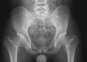
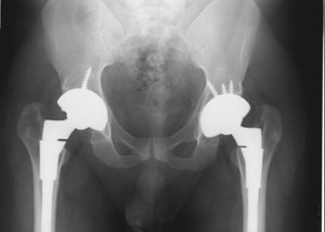
1. Patient “A”: Primary Total Hip Replacement using the ‘muscle- sparing’ (ASI) approach.
 surgery for Primary Total Hip Replacement.")
Severe bi-lateral hip osteoarthritis.
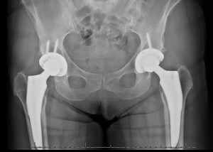
Appearance after replacement of both hips.
Patient “A” was a 64 year old female patient with severe osteoarthritis of both hips. I performed replacement of both her hips, 6 months apart, using the anterior supine, inter-muscular (ASI-muscle sparing) surgical approach. She had an excellent outcome and stated that her hips felt and functioned like her natural hips again.
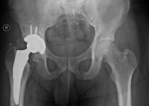
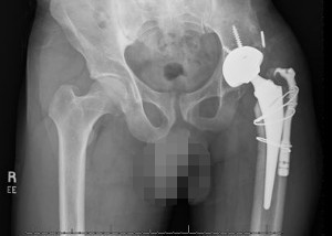
2. Patient “B”: Severe right hip arthritis.
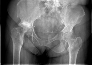
Severe right hip osteoarthritis, with marked pelvic tilt.
After hip right total hip replacement, showing an almost level pelvis.
.This 59 year old woman came to me with severe right hip arthritis with erosion of the acetabulum ( hip socket) and pelvic tilt, resulting from hip contracture. This resulted in a very painful and abnormal gait. After hip replacement, her pain was completely relieved, her pelvis levelled out and she was able to walk with an almost normal gait.
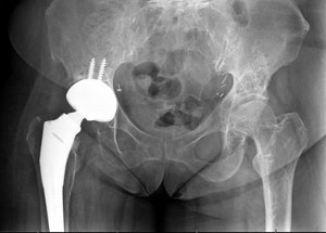
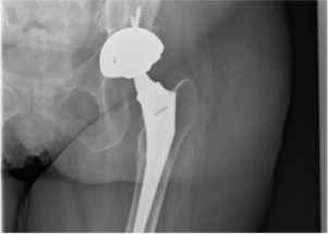
3. Patient “C”: Right hip osteoarthritis with collapse and deformity of the head of femur.
Before hip replacement surgery showing collapse and deformity of head of femur.
After hip replacement surgery.
.Patient “C” was a 62 year old man with severe right hip arthritis with collapse and deformity of the head of femur. Apart from pain and stiffness in his hip, he had marked shortening of his lower extremity. After hip replacement, his pain was relieved, a good range of motion was restored and his leg lengths became equal.
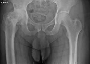
4. Patient “D”: Severe hip arthritis and dislocation of the hip.
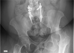
Before hip replacement surgery showing severe arthritis and dislocation of the left hip.
After total hip replacement surgery.
.55 year old patient “D” had partial paralysis of both her legs, after previous spine surgery performed many years earlier at another institution. She also had severe left hip osteoarthritis and dislocation of the left hip and was in constant pain, even when sitting or lying. After hip replacement, her pain was relieved and she was able to sit and lie without pain and was even able to walk short distances with a walker.
5. Patient “E”: Bilateral Hip Replacement for Severe Developmental Hip Dysplasia.
Severe Bilateral hip deformity and arthritis.
After Bilateral hip replacements using special implants.
.This 22 year old man had severe pain in both hips, secondary to arthritis, which was the result of developmental hip deformities. The pediatric orthopaedic surgeons, who had treated him for many years at a renowned pediatric orthopedic hospital, were unable to offer any corrective surgery for his hips. He was referred to me for replacement of both his hips. After I performed these surgeries, using special implants, his hip pain was relieved. He was able to walk with a greatly improved gait and perform his daily activities without any problem.
6. Patient “F”: Conversion of a previous hip fusion to a total hip replacement.
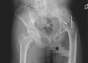
Image of left hip fusion.
Appearance of left hip fusion converted to a total hip replacement.
.Patient “F”, a 48 year old man, came to me with complaints of severe back pain and a stiff left hip. When he was 15 years old, he underwent arthrodesis ( surgical fusion of his left hip) for a neglected traumatic hip dislocation. I converted his fusion to a total hip replacement. This completely relieved his back pain and greatly improved his stance and gait.