
Below is a list of the knee surgeries that I frequently perform:
Sports Medicine
- Arthroscopic- assisted Anterior Cruciate Ligament (ACL) Reconstruction
- Arthroscopic-assisted Posterior Cruciate Ligament (PCL) Reconstruction
- Arthroscopic medial and lateral menisectomy and meniscus repairs
- Arthroscopic treatment of articular cartilage (joint cartilage) loss by microfracture and osteoarticular (cartilage plug) transfer
- Arthroscopic and open treatment of patella instability and dislocations
- Surgical treatment of patella tendinitis
- Repair of quadriceps and patella tendon tears
Joint Replacement:
- Primary Total Replacement
- Partial (Unicompartmental) Knee Replacement
- Revision Replacement
The following X-ray images depict the before and after appearances of KNEE surgeries I have personally performed. In order to protect patients’ identities, their names have not been used and all identifying features have been removed from these images.
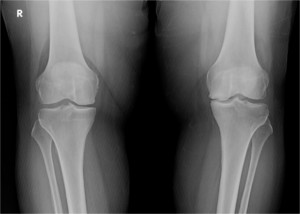
1. Patient “G”: Total Knee Replacement for osteoarthritis of the knee with varus (bow leg) deformity.
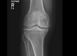
Before Knee Replacement Surgery.
After Knee Replacement Surgery.
Patient “G” was a 68 year old man with severe and disabling right knee pain. His pre-operative X-rays shows advanced osteoarthritis of the knee with a varus ( bow leg) deformity. The post op X-ray shows the knee replacement in place with the normal knee alignment restored.
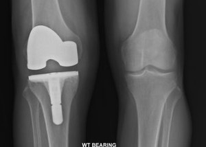
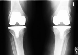
2. Patient “H” : Total Knee Replacement for osteoarthritis with severe varus (bow leg) deformity.
Arthritis with severe varus (bow leg) deformity.
Post-op image of total knee replacement for arthritis and varus (bow leg deformity).
Correction of knee deformities, whether varus (bow leg) or valgus ( knock knee), is an important and integral part of total knee replacement done for knee arthritis. This is shown in the pre and post op X-ray images of patient “H”, a 72 year old man who had severe bow leg deformity. Also seen on the early post-op X-ray image of patient “H” ‘s knee are the metal skin staples used for closure of his surgical wound.
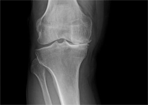
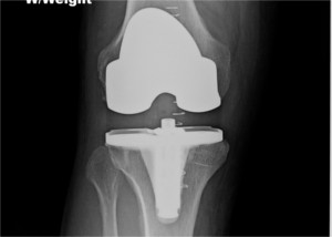
3. Patient “I” : Partial Knee Replacement for unicompartmental arthritis of the knee:
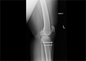
Before surgery for arthritis of medial (inner side) compartment of knee.
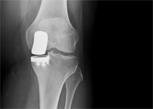
After partial knee replacement surgery for arthritis of medial ( inner side) compartment of knee.
.When a patient has severe arthritis involving only the medial (inner side) or lateral (outer side) compartment of the knee, a partial knee replacement is an excellent operation which is less invasive and involves an easier rehabilitation. This is shown in the above post-op X-ray of patient “I”, a 52 year old woman who had failed to respond to all non-surgical treatments.
4. Patient “J”: Revision Knee Replacements, using special implants, for painful loosening of previous knee replacements.
Before Revision Surgery, showing old knee implants from 14 yrs. ago.
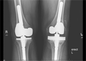
After Revision surgery of both knees.
By age 73, patient “J” had already had both knees replaced 14 years earlier at another institution. He had pain in his knees because of loosening of the components. The post-op X-ray shows the revision (re-do) prosthesis. Note the long stems used for stability to compensate for bone loss and ligament damage from his loosened initial knee replacements.
5. Patient “K”: Patello- Femoral (Knee Cap Joint) re-alignment surgery for patello-femural arthritis with mal-alignment.
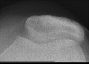
Before surgery showing severe patello-femural arthritis.
Post op X-ray showing central position of knee cap and restored joint space.
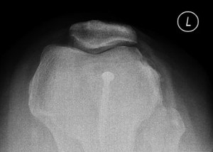
Post op X-ray showing central placement of patella (knee cap).
This 46 year old lady had severe left knee pain because of patella-femoral ( knee cap joint) arthritis with associated patella mal- tracking (mis- alignment). Her pain was completely relieved by a Fulkerson Osteotomy. This operation involves cutting and moving the bone at the sight of the patellar tendon attachment. Note the central position of the knee cap and the restored joint space seen in the post- op X-ray.